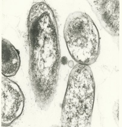
Chronic infections can persist for various reasons. A weakened host immune system may provide a suitable environment for bacteria to thrive long-term. Likewise, re-infections could take place due to shared habitats between host and vector. The ability for tick-borne pathogens, like Bartonella and Borrelia, to form biofilms is another possibility that clinicians and researchers are actively exploring.
What is a “biofilm”?
A biofilm is composed of microbes that colonize and aggregate on a moist surface. The colony can be composed of a single bacterial species, or a heterogenous mixture of species that include microorganisms such as fungi and/or protozoa. The plaque that builds up on teeth and leads to painful cavities is one example of a biofilm. Bacteria can also collect on surfaces of the body that are not so easily monitored, such as the vascular system.
Biofilm formation typically begins when cells encounter stressors that trigger production of specialized attachment proteins and structures. The structures, called pili, allow for initial attachment to a moist surface while the proteins, or adhesins, make the cells “sticky”.
Once adhered to a suitable surface the cell begins secreting a slimy material, called extracellular polymeric substance, which causes an accumulation of cells over time. As the colony develops an outer layer composed of sugars and minerals is created which allows for the cells inside to interact.
The resulting colony begins to behave a bit like a multicellular organism. Despite the outer layer offering a protective barrier, it is difficult for internal cells to access nutrients that are necessary for cellular division. Cells must communicate via signaling pathways called quorum sensing to overcome this challenge.
Some cells will adopt specialized functions to keep the entire unit alive. An example is the creation of channels that can facilitate transport of water and nutrients Over time, biofilms cause damage to the attached surface and individual cells can leave the colony to create colonies elsewhere.
The main reasons for biofilm formation are survival strategies during stressful events like:
- Reduced needs when resources are limited– Nutrient deprivation can lead to aggregation of cells because less energy is needed as a group.
- Protection from antibiotics– The outer layer protects the group from antibiotics penetrating cells that are located inside. Some cells in the colony may also absorb additional antibiotics in a self-sacrifice role that protects the colony. Research shows that antibiotics can be up to 80% less effective against biofilms.
- Protection from immune system– Like antibiotics, host immune cells cannot easily invade the biofilm because normal surface proteins on the bacteria are concealed by the outer layer.
- Combined resources with other species– In one research example, Borrelia lymphocytoma samples contained DNA from Chlamydia species in the biofilm. Researchers hypothesized that the bacteria formed a symbiotic relationship in order to survive long-term.
Clinical Relevance
Biofilm formation is a well-known issue for medical equipment in hospital settings. Bacteria, such as Pseudomonas species, colonize moist tracheal tubes and then cause human infections when used again. Despite the hospital using proper cleaning protocols between procedures, bacteria remain because antimicrobials are not effective. Furthermore, the infections that result are often antibiotic resistant.
Bartonella and Borrelia species have both been shown to form biofilms in both laboratory settings as well as human clinical samples. A large amount of research is delving into how biofilm formation occurs in humans and whether chronic infections can be attributed to it.
B. henselae and B. quintana form biofilms on heart valves in cases of Bartonella endocarditis. Studies show that these cases may be linked to patients that already had heart valve disease, so the bacteria are able to easily colonize the damaged tissue. However, research has shown these species can produce large amounts of adhesins that promote biofilm formation. Therefore, when Bartonella species are present, they can “stick” to the fibrous heart valve tissue and slowly aggregate.
Conclusion
When bacteria form biofilms, they become more than the sum of their parts. This impacts human health from sanitation to infection treatment. As one more piece of the puzzle that suggests medicine is moving beyond the “one pathogen, one disease model,” researchers are now looking at how biofilms and their combined host protein and multi-species bacterial makeup affect treatment options.
References
Kumar, A. et al. (2017). Biofilms: Survival and defense strategy for pathogens. International Journal of Microbiology, 307(8), 481-489. doi:10.1016/j.ijmm.2017.09.016 https://www.researchgate.net/publication/319965153_Biofilms_Survival_and_defense_strategy_for_pathogens
Okaro, U. et al. (2017). Bartonella species, an emerging cause of blood-culture-negative endocarditis. Clinical Microbiology Reviews, 30(3), 709-746. doi:10.1128/CMR.00013-17 https://www.ncbi.nlm.nih.gov/pubmed/28490579
Sapi, E. et al. (2019). Borrelia and Chlamydia can form mixed biofilms in infected human skin tissues. European Journal of Microbiology and Immunology, 9(2), 46-55. doi:10.1556/1886.2019.00003 https://www.ncbi.nlm.nih.gov/pubmed/31223496
Timmaraju, V. A. et al. (2015). Biofilm formation by Borrelia burgdorferi sensu lato. FEMS Microbiology Letters, 362(15), fnv120. doi:10.1093/femsle/fnv120 https://www.ncbi.nlm.nih.gov/pubmed/26208529
Di Domenico, E. G. et al. (2018). The emerging role of microbial biofilm in Lyme neuroborreliosis. Frontiers in Neurology, 9, 1048. doi:10.3389/fneur.2018.01048 https://www.ncbi.nlm.nih.gov/pubmed/30559713
Okaro, U. et al. (2019). The trimeric autotransporter adhesin BadA is required for in vitro biofilm formation by Bartonella henselae. NPJ Biofilms and Microbiomes, 5, 10. doi:10.1038/s41522-019-0083-8 https://www.ncbi.nlm.nih.gov/pubmed/30886729
Snoussi, M. et al. (2018). Heterogeneous absorption of antimicrobial peptide LL37 in Escherichia coli cells enhances population survivability. eLife.2018,7:e38174. doi:10.7554/eLife.38174 https://elifesciences.org/articles/38174


